Tight Laminar Connection – Founder Prevention

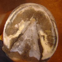
Healthy laminae are vital  in preventing founder. The health of the laminae is determined in part by their length. The shorter they are, the more tightly they connect the coffin bone to the inside of the hoof wall, because they are not ‘stretched out’.  Their distance is measured horizontally from the coffin bone to the inside of the hoof wall. On the sole view, they appear as a narrow, tight, healthy white line with no separation.  

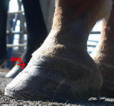
The photos  in this progression essentially speak for themselves, and are used to illustrate the change in the laminar connection, as well as the angle of growth, once a correct trim is undertaken. Equally obvious results are evident in horses that are already barefoot but were not correctly trimmed, and examples of these will be posted in future entries. (Reader contributions as always are welcome).

1aug9bfm.jpg August

The horse was ‘rescued’ from a hack line by a new owner at this point. The horse was bruising himself with the shoes due to the overgrown feet.


2sep06m2.jpg September

One month after the shoes were removed, the horse has largely self-trimmed its feet to this profile with the exception of the toe being rasped a little by hand. The new laminar growth has come in so much tighter (closer to the coffin bone) that the hoof wall has ‘separated’ away at the line of new growth and looks like a crack. The second half of the new growth has come in at a more correct angle as the hoof started correcting itself.  


3oct15m.jpgOctober

The new growth has progressed almost halfway down the hoof and continues to come in at a steeper angle, closer and tighter to the coffin bone.


4nov1m-3.jpgNovember

 A small amount of old growth remains, projecting beyond the new growth.


5nov28m.jpg December

The growth is now all at the same angle and the ‘cracked’ hoof wall has almost grown out.


 The pastern angle remains steep as a result of joint adaptation (from the long term incorrect hoof form visible in August), rather than heel pain.

Turtleback Zoo of Essex County, NJ Opts For Natural Hoofcare

sign1.jpg


dantethedraft.jpgDante is a 9 year old rescued Belgian Draft Horse. He was once an Amish work horse.  Upon entering the zoo he was lame regularly and needed bute often for pain control.  The zoo’s farrier could not control him even with sedation, and ended up only rasping the tops of his feet for months.  Finally, a natural barefoot hoofcare professional was sought out, in a last ditch effort to get him sound and controllable before giving up and sending him away again.  What a turn around!!!  Dante has been sound since his first trim.  The trim addressed his overgrown bars, frog, and major flaring, and took almost 3 hours!  Four months later, he is getting better and dantethedraft-21.jpgbetter.  He no longer needs sedation, thanks to time and patience, but mostly due to some natural horsemanship techniques that have encouraged him to give his foot and stand quietly.  Dante is a pleasure to work with now and the zoo staff is amazed at how far he’s come.  He is one of the most popular animals at the zoo.  Check him out one day!!

www.turtlebackzoo.com

dante.jpg

Description of the Horse’s Hoof & Corium

corium3corium2

Laminae, Laminar Corium, Coffin Bone Corium

Laminae, Laminar Corium, Coffin Bone Corium

Written By Odette Suter, DVM 

Laminae are composed of a sensitive and insensitive part. The sensitive part is also known as the laminar corium and the insensitive laminae is also called laminar horn and is produced by the laminar corium. The corium is a form of epithelium. Epithelial tissue is composed of one or more layers of cells that line surfaces of organs (skin, GI tract, lungs, etc.), blood vessels, lymph vessels, etc. The functions of epithelial tissue are
protection, absorption, diffusion as well as excretion of waste products. In the case of the hoof this ‘waste’ is horn (protein).

Dr. Odette Suter
www.odettesuter.com

animal communication
equine hoof rehab
spiritual consulting


All photos courtesy Kentucky Horsehoeing School

Filly with Club Foot

rfsm.jpgThis is a 9 month old filly whose right front foot is clubby. It has been shod on the recommendation of several vets and farriers who believe the tendon is ‘tight’ (despite no evidence to indicate this), and shoeing it and gradually lowering the heel is expected to correct this situation.

The toe wall has begun to bulge out,  possibly  as a result of the shoe ‘holding’ the bottom of the foot while the rest of the foot tries to grow outward.


lfsm.jpg The left front leg is shown for comparison. Note the differences in the angle of the long pastern bone (P1)  in relation to the  short pastern bone (P2)


Continue reading

Relationship of Hoof Form to Metabolism, Cresty Necks & Founder

”Insulin Resistance” is a trendy diagnosis made by lay people with regards to their own horses, based on information they glean from the internet. It is suspected as a cause in laminitis and founder, and horses are removed from most or all pasture, depriving them of their natural grazing needs in an attempt to remove sugars from the diet. I believe it is possible that this actually can cause the horse to experience even more stress and thus puts in motion a vicious cycle.Below are some excerpts from an article in The Horse, in which Tia Nelson DVM, discusses her experience in reducing signs of insulin resistance (such as cresty necks) in some horses, simply by improving their hoof form.

As she says, it is important to manage horses’ diets, especially easy keepers, but it can be very helpful to eliminate poor hoof form as a contributing factor. There is more on Insulin Resistance in a study posted on this blog, linked on this page here

Read the whole article here

Continue reading

Holiday Greetings

kel.jpg

from our rescue case (right) and his barefoot companion, who appears to be thriving in his new digs in Nevada.  He moved there with his owner after we helped rehab him (as you read in his case history in a previous thread).  At the time he was reclaimed by his owner, it was questionable whether he would live; today – he is modelling a silly reindeer antler and Santa hat. Life is good!

Best wishes to all in 2007.

Body Posture and Hoof Form

How a Horse’s Stance is Affected by The Feet and their Balance

Shown here are the horse’s front feet.

right-front-sole.jpg    also-right-f-sole.jpg

Right Front, two views

 left-front-sole1.jpg    

Left Front

Based on the trim and balance of the two front feet, how would you expect the horse to stand?

  1. Standing Squarely
  2. Right Foot Placed Behind Left
  3. Left Foot Placed Behind Right
  4. Both Front Feet Placed Behind Vertical

To see the answer,   Continue reading

Foot Abscesses

  abs2.jpg 


Heel Bulb Abscess


 abs1.jpg Sole Abscess – between frog & bar on left

above two photos courtesy Amy Dickson, Kamloops BC


 Sole/HeelAbscess

solarheelabs.jpg

While it was ‘active’, this abscess created a pocket of fluid that impinged on the blood vessels in the solar corium, bruising it (visible as the pink edge around the outer edge of the abscess). 


Continue reading