Founder – Laminar Wedge

The Laminar Wedge

is the space in the front of the hoof at the toe, where stretched and torn laminae have caused the coffin bone to rotate away from the hoof wall.   The space between coffin bone and hoof wall has widened at the bottom, relative to the top, and has filled with wound secretion, blood and torn and dead laminae.

rflamwedge.jpg

From the Front

The ‘dead’ laminae are black.  

lamwedgetop.jpg

From the Top

lamwegeside.jpg

From the side. The tip of the toe no longer contacts the ground.

(the photos are of both front feet, both of which foundered).

Overgrown Bar & Sole, Abscess & Bruise

 The horse has relatively flat soles, so the buildup of excess sole was especially harmful and painful.

Right Front Foot


 rfsolebf.jpg    rfsoleaf.jpg

Before                                                                               After

Bruises were revealed underneath the excess sole immediately after it was trimmed away.

Left Front Foot

            beforelfa.jpg               lfsoleaf.jpg

(click on thumbnail for larger image)

Before                                                   After

This foot had already developed abscesses underneath the excess sole, corresponding exactly to the outline of the sole ‘lumps’, visible in the small thumbnail to the left.

Sidebone Xrays

Left Front

 lf-ap.jpg  

Right Front

 rf-ap.jpg

Here is an example of a horse with sidebone on both front feet. The lateral cartilages have ossified into what looks literally like a bone on the sides. The parts that appear ‘broken’ are most likely not fully ossified  yet.

The plainly prominent and deep collateral grooves can prevent flexing of the foot which further prevents shock absorption, which can contribute to the formation of sidebone.

Uneven Hoof Walls

This is a Left Front foot with the inside wall curving to the inside, in a bell-shaped fashion (red dotted line Fig. 2).  This is being caused in part by the sole at the inside toe which is  too high (Fig. 1) as well as the toe too long.

This young mustang, never shod, has very thick and extremely hard walls which do not break off with wear but continue growing in a distorted way, and altering the horse’s stance.

1alfbefmu.jpg    4lffrontmu.jpg

Fig. 1                                                          Fig. 2

The trimming treats this condition similarly to a flare but also lowers the toe on the inside (sole is removed). So, there are cases when sole forward of the frog does need to be rasped. The trimming is complicated by the inside-growing direction the walls have adopted. Now that the inside wall is growing more correctly, the outside wall needs to begin to start growing in a more outwardly direction (more visible in Fig 3 due to the angle of the photograph). Advanced trimming techniques such as floating the heel and the diagonal toe will allow this to occur.

2alfheelsnov1mu.jpg   5lffrontoctmu2.jpg

Fig. 3                                                     Fig. 4


7lffrontwavesepmu.jpg

Fig. 5

A photo from an early trim shows how all the pressure was  put on the inside wall by the long toe, with all the compression rings on the inside only.
 

Overgrown Bars – ‘False Sole’, ‘Double Sole’

Here is an example of overgrown bar that has migrated forward onto the sole creating the appearance of a ‘false’ or ‘double’ sole.  The material is not merged with the sole and is distinctly different in appearance and texture.

   right-fore-solar-initial.jpg    right-fore-lateral-initial.jpg 

After 5 months of trimming:

right-fore-solar-5-months-later.jpg

The bars are brought back to their correct position ending alongside the collateral grooves and approximately to the midpoint of the frog. Also note the expansion of the contracted heels.

These pictures are courtesy of a barefoot trimmer in the Midwest.

Overgrown Bars

The horse in question is a 30+ (?) yo neglected (starvation) rescue case.

Overgrown bars can lead to a variety of problems, such as exerting pressure on the hoof wall which causes it to crack.

jerry-crack1.jpg

The Right Hind hoof has a crack starting at the lateral toe quarter which began with an abscess. (As seen here).

jerry-bar1.jpg   jerry-sole-aft1.jpg

 Before trim                                            After trim

Both bars are too long, grown above the height of the frog preventing it from being weightbearing. The lateral bar has extended almost to the frog’s apex and overlaid onto the sole. At the green arrow it is raised above the sole by at least 1/4″.  The furthest point of the bar (where the blue arrow starts), exerts pressure on the wall when the foot is weightbearing, in the direction of that arrow and splits the wall apart.  The underrun heels also contribute to the problem. To correct the problem, the bars have been shortened and brought back approximately to the frog’s midpoint, the heels have been lowered and brought back to the widest point of the frog, and the toe has been backed up. These changes will allow proper forces to be exerted on the hoof capsule allowing the crack to grow out.

Founder and Laminitis – Effect of Hoof Form

 Founder & Laminitis (Under Construction)

The following diagram comes from the May ’99 issue of Practical Horseman:

laminitis2.jpg

This is a good depiction showing the progression from a healthy foot to a rotated one.  However, the heels which give us most of the information about the possibility of rotation, have been obscured.

The important information with respect to rotation, laminitis, and founder can be found in the thickness of the sensitive laminae, as well as the orientation of the coffin bone relative to them.  

laminitis3a.jpg                 sunny3.jpg

1.  Healthy Foot.  The sensitive laminae (pink) are close, tight, and parallel to the insensitive laminae (white line).  The actual foot resembles the shape of the healthy, diagrammed one.  With its low heel, the coffin bone is parallel to the toe wall which is the surest way to ensure a healthy white line and healthy, tight, laminae that can solidly suspend the coffin bone and prevent founder espisode.

laminitis4a.jpg                   logan2.JPG

2.  Mildly Pathological Foot.  The wall of the coffin bone is no longer parallel to the hoof wall which causes the laminae to stretch at the bottom and begin to adopt a ‘wedge-shape’: narrower at the top at wider at the bottom. This puts constant stress on the overstretched laminae towards the bottom.

laminitis5a.jpg                   crimmy2.JPG

3.  Severe Pathology. Founder – Rotation with Separation.  The ‘wedge’ is extreme now, with the laminae much longer at the bottom than the top.  The bone is far out of parallel with the hoof wall.  The constant downward stress and pull on the lower part of the laminae has caused them to fail, allowing the suspension of the bone to the inside of the hoof wall to be lost and the bone to rotate. 

Collateral Grooves and Bars

Below is an Xray of a Right Front hoof.  The right side is the medial side, the left is the lateral one. (The view is from the front).

rf-ap-bar3.jpg

The two views are the same, one shown un-marked up for clarity.

rf-ap-bar2.jpg

 The tops of the collateral grooves are indicated with red marks.  The depth of the collateral groove is determined by the height (or length) of the bar, which is not visible on xray. The collateral groove on the medial side is very deep, much more so than the lateral side. It appears to be projecting up into the bottom of the coffin bone which is probably very painful, since this area is full of sensitive tissue.  The bar and collateral groove has pushed up the inside side of the foot, displacing the balance of the foot.   The inside wall is much higher and longer than the outside. The hairline and top of the wall/coronary band are indicated with the red arrows. The two points should be straight across, parallel with the ground. Another detrimental effect of this deep collateral groove is the displacement of the P3/P2 joint. The joint space is narrower on the outside, wider on the inside.  This can lead to ossifications and arthritic problems such as sidebone and ringbone.

Lowering the bar on the inside to the same length as the outside will correct these balance problems.

Tight Laminar Connection – Founder Prevention

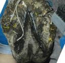
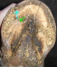
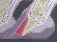
Healthy laminae are vital  in preventing founder. The health of the laminae is determined in part by their length. The shorter they are, the more tightly they connect the coffin bone to the inside of the hoof wall, because they are not ‘stretched out’.  Their distance is measured horizontally from the coffin bone to the inside of the hoof wall. On the sole view, they appear as a narrow, tight, healthy white line with no separation.  

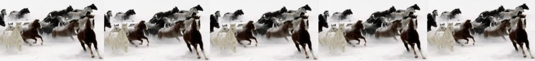
The photos  in this progression essentially speak for themselves, and are used to illustrate the change in the laminar connection, as well as the angle of growth, once a correct trim is undertaken. Equally obvious results are evident in horses that are already barefoot but were not correctly trimmed, and examples of these will be posted in future entries. (Reader contributions as always are welcome).
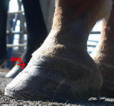
1aug9bfm.jpg August

The horse was ‘rescued’ from a hack line by a new owner at this point. The horse was bruising himself with the shoes due to the overgrown feet.
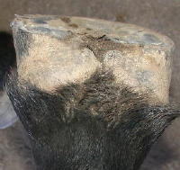


2sep06m2.jpg September

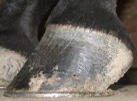
One month after the shoes were removed, the horse has largely self-trimmed its feet to this profile with the exception of the toe being rasped a little by hand. The new laminar growth has come in so much tighter (closer to the coffin bone) that the hoof wall has ‘separated’ away at the line of new growth and looks like a crack. The second half of the new growth has come in at a more correct angle as the hoof started correcting itself.  


3oct15m.jpgOctober

The new growth has progressed almost halfway down the hoof and continues to come in at a steeper angle, closer and tighter to the coffin bone.


4nov1m-3.jpgNovember

 A small amount of old growth remains, projecting beyond the new growth.


5nov28m.jpg December

The growth is now all at the same angle and the ‘cracked’ hoof wall has almost grown out.


 The pastern angle remains steep as a result of joint adaptation (from the long term incorrect hoof form visible in August), rather than heel pain.