Cross Section Through Shod Hoof

This is an image of a cross section through a hoof with the shoe still attached and showing how the nail goes through the foot into the white line.  It appeared originally in The Hoof Blog, in a post discussing the use of MRI’s for diagnostic purposes and the proper removal of shoes

http://hoofcare.blogspot.com/2011/02/no-farrier-no-mri-diagnostic-imaging.html

This is a fascinating, rarely seen view of the shoe and nails penetrating the hoof.  It is remarkable how close the nail is to the edge of the coffin bone.  Click on the image for a larger view.

Hoof Cross Section With Shoe and Nails Attached

Hoof Cross Section With Shoe and Nails Attached

Founder – Laminar Wedge Xrays

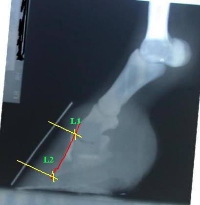
The following xrays depict radiographically the laminar wedge as well as the rotation characteristic of founder. They correspond to the photographs of the laminar wedge in the previous post.

lfmu2.jpg

The horse was diagnosed with rotation of 12 degrees. The space between the laminae at the top of the hoof (L1) is smaller than the space at the bottom (L2). These two measurements should be the same, or stated another way the wall of the Coffin Bone (red line) should be parallel to the hoof wall (metal marker seen in gray). When the space at the bottom is greater this indicates the laminae have given way and the bone has begun to rotate, or founder. The wide space at the bottom, L2, is now full of torn and dead laminae, inflammation and blood, and this is what becomes the laminar wedge.

The importance of correct laminar spacing (he calls it the Horn-Laminar zone) is discussed by Ric Redden here

Here is an umarked Xray of the same foot:

lfmu.jpg

Click on thumbnail for larger image.

Founder – Laminar Wedge

The Laminar Wedge

is the space in the front of the hoof at the toe, where stretched and torn laminae have caused the coffin bone to rotate away from the hoof wall.   The space between coffin bone and hoof wall has widened at the bottom, relative to the top, and has filled with wound secretion, blood and torn and dead laminae.

rflamwedge.jpg

From the Front

The ‘dead’ laminae are black.  

lamwedgetop.jpg

From the Top

lamwegeside.jpg

From the side. The tip of the toe no longer contacts the ground.

(the photos are of both front feet, both of which foundered).

Sidebone Xrays

Left Front

 lf-ap.jpg  

Right Front

 rf-ap.jpg

Here is an example of a horse with sidebone on both front feet. The lateral cartilages have ossified into what looks literally like a bone on the sides. The parts that appear ‘broken’ are most likely not fully ossified  yet.

The plainly prominent and deep collateral grooves can prevent flexing of the foot which further prevents shock absorption, which can contribute to the formation of sidebone.

Overgrown Bars – ‘False Sole’, ‘Double Sole’

Here is an example of overgrown bar that has migrated forward onto the sole creating the appearance of a ‘false’ or ‘double’ sole.  The material is not merged with the sole and is distinctly different in appearance and texture.

   right-fore-solar-initial.jpg    right-fore-lateral-initial.jpg 

After 5 months of trimming:

right-fore-solar-5-months-later.jpg

The bars are brought back to their correct position ending alongside the collateral grooves and approximately to the midpoint of the frog. Also note the expansion of the contracted heels.

These pictures are courtesy of a barefoot trimmer in the Midwest.

Collateral Grooves and Bars

Below is an Xray of a Right Front hoof.  The right side is the medial side, the left is the lateral one. (The view is from the front).

rf-ap-bar3.jpg

The two views are the same, one shown un-marked up for clarity.

rf-ap-bar2.jpg

 The tops of the collateral grooves are indicated with red marks.  The depth of the collateral groove is determined by the height (or length) of the bar, which is not visible on xray. The collateral groove on the medial side is very deep, much more so than the lateral side. It appears to be projecting up into the bottom of the coffin bone which is probably very painful, since this area is full of sensitive tissue.  The bar and collateral groove has pushed up the inside side of the foot, displacing the balance of the foot.   The inside wall is much higher and longer than the outside. The hairline and top of the wall/coronary band are indicated with the red arrows. The two points should be straight across, parallel with the ground. Another detrimental effect of this deep collateral groove is the displacement of the P3/P2 joint. The joint space is narrower on the outside, wider on the inside.  This can lead to ossifications and arthritic problems such as sidebone and ringbone.

Lowering the bar on the inside to the same length as the outside will correct these balance problems.

Description of the Horse’s Hoof & Corium

corium3corium2

Laminae, Laminar Corium, Coffin Bone Corium

Laminae, Laminar Corium, Coffin Bone Corium

Written By Odette Suter, DVM 

Laminae are composed of a sensitive and insensitive part. The sensitive part is also known as the laminar corium and the insensitive laminae is also called laminar horn and is produced by the laminar corium. The corium is a form of epithelium. Epithelial tissue is composed of one or more layers of cells that line surfaces of organs (skin, GI tract, lungs, etc.), blood vessels, lymph vessels, etc. The functions of epithelial tissue are
protection, absorption, diffusion as well as excretion of waste products. In the case of the hoof this ‘waste’ is horn (protein).

Dr. Odette Suter
www.odettesuter.com

animal communication
equine hoof rehab
spiritual consulting


All photos courtesy Kentucky Horsehoeing School