These Are Not Reasons To Shoe A Horse, Young or Old

horse1Interesting article from The Horse, addressing the question whether older horses need to be shod. I find these to be the most frustrating sort of articles to read. The author comes to the conclusion that some horses ‘just need shoes’ (as so many so often “just” do). But, not only are her ‘reasons’ for shoeing flawed, nothing in them or the article addresses the specific issue of an older horse needing shoes. And think about it – what about an older horse would make his hoofcare needs any different from a younger one’s? (Answer: nothing) Only thing I can think of is that they’re assuming an older horse’s workload is reduced and thus the need for shoeing. But the article doesn’t go into that, discussing active horses.


Although he has pretty decent feet for a Thoroughbred, it was pretty clear he needed some extra support to successfully navigate the varied terrain on which eventing takes place, based on his short and choppy stride coupled with the fact that his front hooves started chipping like crazy once he started jumping. Our farrier tacked on a pair of front shoes and things have been going well. Continue reading

Shod To Barefoot Transition – Long Toe Low Heel

A 5 yo Hanoverian/TB X gelding

Shoes were removed in December.  The images represent his progress over the course of the next 5 months.

Left Front Foot

Dec082lf      Mar09lf

Fig. 1                                                              Fig. 2

Continue reading

Navicular, a.k.a ‘Caudal Heel Pain Syndrome’

What a Little Good Trimming Can Do

dscn4089.jpg

The horse in question, an 18 yo Appaloosa was retired from showing because of the non-specific diagnosis of caudal heel pain syndrome.  Xrays confirmed the presence of ‘changes’ that were attributed to his discomfort.  He was shod according to proper conventional veterinary standards for navicular, which did help to make him comfortable, but he still seemed stiff, definitely not agile, and, at the bottom of the pecking order would allow himself to be cornered and bullied rather than try and run away.

Right Front Leg Lateral View

1may07b.jpg           2may07b.jpg         3my07b.jpg

a.  With shoes                          b.  Shoes Just Removed            c.  First Trim

Continue reading

Ringbone?

OR POOR HOOF FORM? 

The Morgan mare is believed to be about 15 yo.  She was found at auction in MA. Due to her severe lameness (grade5/5 at a the walk), no one wanted her and for several weeks she wasted in the auction pens.  She was shod, but according to the sellers it did not help, and even with Banamine she was completely lame on some days.  She was in danger of getting picked up by a slaughter-bound truck when by chance the current owner found her and purchased her for $400. 

(Click on thumbnails for larger views).

  lily002.jpg                      lily-fink0003.jpg

        Left Hind AP                            Left Hind Lateral

This horse clearly has a very advanced case of high, apparently articular, ringbone.  According to the veterinary diagnosis, it was the most severe case ever seen by that vet and the horse would never be sound for riding.
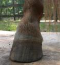
lhringbone.JPG        lhbfrb.JPG   rlh.JPG

Left Hind

The ringbone is clearly visible even without radiographs and the mare frequently favored the Left Hind.

Moving on from what is visible on radiographs, the obvious confronts the viewer: the horse’s hoof form is terrible and overgrown, the result of neglect or ignorance. There is certainly more than enough cause here for lameness of some degree.
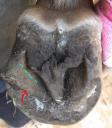
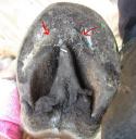
2lhsolebf.JPG      1lhsolebf.JPG         3lhsole.JPG

LH before, fig. 1                    LH before, fig. 2             LH after, fig. 3

The bars on the Left Hind are clearly overgrown to the point where they are actually above not only the level of the sole but the wall as well, meaning the bar would be the first structure to bear the horse’s weight, upon weightbearing rather than the walls and sole.  Since the foot is somewhat contracted and the wall and bar material are very hard (as is typical in Morgans), the bars are not folding over onto the sole, the effect for the horse being like stepping onto the dull edge of a knife with each step.  No wonder she refused to put any weight onto that foot.

Fig. 1 shows the edge of the too-long bar (red arrow) as well as the desired location of the bar (blue dashed line). Fig. 2 shows the bar grown all the way around the apex of the frog (red arrows), also a source for pain. Fig. 3 shows the bars lowered and removed from the sole.  After this trim the mare was much more willing to stand on this foot but was still lame on turns.

Having become more comfortable on the LH, she now exhibited more clearly lameness on the Right Front and is seen holding that foot behind her, a sign of pain.

1body.JPG

Further investigation revealed deeply imbedded bar on the RF front, which when removed, produced immediate improved soundness.

Right Front
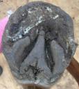
1rfsolebf.JPG                        3rfsolaf.JPG   

Before                                                       After

 2rflatbf.JPG                   4rflataf2.JPG

Before                                                      After

Update:  The mare has been under the new owner’s care for about six months now.   After her first few trims she was able to place weight on her feet and move comfortably, so she was started on trail rides of increasing duration, sometimes as much as 4 hours long.  After the very longest rides she would show some signs of discomfort in her hind legs, which presumably was the articular deposits being worn away from the hours of movement. (This will be confirmed in the coming months with new X-rays).   But evn this discomfort is no longer present. It is apparent that the obvious pain and inability to place weight on the Left Hind was orginating from the large overgrown bar seen from the underside on the lateral side of the foot, even though this was never observed in the lameness diagnosis.  The lameness was all attributed to the ringbone.  She requires no boots on every kind of footing in the park where she trail rides. 

Founder – Recovery

4 yo Shetland Pony Mare

The pony had not been trimmed much until the time she foundered, and grazed on lush grass while under the care of the previous owner, resulting in a combination of probable metabolic and mechanical founder. Her X-rays and laminar wedge closeups appear in the posts below.

Ruby Standing

A tight regimen of frequent trimming as well as limited access to grass (using a muzzle) has been implemented, resulting in improved hoof form and a healthier body weight.  The trimming focused on lowering the heels and backing up the toes, realigning the coffin bone parallel to the toe wall, as well as bringing it closer to a ground parallel orientation. The parallel hoof wall/coffin bone is a primary factor in the prevention of founder.

BEFORE

rfbefdecember-2.jpg   solebf.jpg

Somewhat difficult to see in the grass, but this is where the corrective trimming started, with high heels and very long toes.  The red arrows at the toe show imminent coffin bone protrusion, along with a wide gulf  separation between its edge and the wall. The bar, (red arrow), level with the frog, is high.

rfsole.jpg       rfsole0307.jpg       rfsolebl.jpg

1 mo.                                  3 mos.                                    6 mos.

As the hoof wall grows down, the separation (all the way around the edge of the hoof) diminishes, and the white line becomes healthy and tight, enabling it to suspend the coffin bone in the hoof capsule.

 rflat.jpg                                              rflatbl.jpg

1 mo.                                                                                   6 mos.

By 6 months most of the hoof wall has grown down with less prominent rings. The remaining separation at the toe (red arrows) corresponds to the separation on the sole at the same time frame and will grow out in another month or so.

Founder – Laminar Wedge

The Laminar Wedge

is the space in the front of the hoof at the toe, where stretched and torn laminae have caused the coffin bone to rotate away from the hoof wall.   The space between coffin bone and hoof wall has widened at the bottom, relative to the top, and has filled with wound secretion, blood and torn and dead laminae.

rflamwedge.jpg

From the Front

The ‘dead’ laminae are black.  

lamwedgetop.jpg

From the Top

lamwegeside.jpg

From the side. The tip of the toe no longer contacts the ground.

(the photos are of both front feet, both of which foundered).

Uneven Hoof Walls

This is a Left Front foot with the inside wall curving to the inside, in a bell-shaped fashion (red dotted line Fig. 2).  This is being caused in part by the sole at the inside toe which is  too high (Fig. 1) as well as the toe too long.

This young mustang, never shod, has very thick and extremely hard walls which do not break off with wear but continue growing in a distorted way, and altering the horse’s stance.

1alfbefmu.jpg    4lffrontmu.jpg

Fig. 1                                                          Fig. 2

The trimming treats this condition similarly to a flare but also lowers the toe on the inside (sole is removed). So, there are cases when sole forward of the frog does need to be rasped. The trimming is complicated by the inside-growing direction the walls have adopted. Now that the inside wall is growing more correctly, the outside wall needs to begin to start growing in a more outwardly direction (more visible in Fig 3 due to the angle of the photograph). Advanced trimming techniques such as floating the heel and the diagonal toe will allow this to occur.

2alfheelsnov1mu.jpg   5lffrontoctmu2.jpg

Fig. 3                                                     Fig. 4


7lffrontwavesepmu.jpg

Fig. 5

A photo from an early trim shows how all the pressure was  put on the inside wall by the long toe, with all the compression rings on the inside only.
 

Tight Laminar Connection – Founder Prevention

Healthy laminae are vital  in preventing founder. The health of the laminae is determined in part by their length. The shorter they are, the more tightly they connect the coffin bone to the inside of the hoof wall, because they are not ‘stretched out’.  Their distance is measured horizontally from the coffin bone to the inside of the hoof wall. On the sole view, they appear as a narrow, tight, healthy white line with no separation.  

The photos  in this progression essentially speak for themselves, and are used to illustrate the change in the laminar connection, as well as the angle of growth, once a correct trim is undertaken. Equally obvious results are evident in horses that are already barefoot but were not correctly trimmed, and examples of these will be posted in future entries. (Reader contributions as always are welcome).

1aug9bfm.jpg August

The horse was ‘rescued’ from a hack line by a new owner at this point. The horse was bruising himself with the shoes due to the overgrown feet.


2sep06m2.jpg September

One month after the shoes were removed, the horse has largely self-trimmed its feet to this profile with the exception of the toe being rasped a little by hand. The new laminar growth has come in so much tighter (closer to the coffin bone) that the hoof wall has ‘separated’ away at the line of new growth and looks like a crack. The second half of the new growth has come in at a more correct angle as the hoof started correcting itself.  


3oct15m.jpgOctober

The new growth has progressed almost halfway down the hoof and continues to come in at a steeper angle, closer and tighter to the coffin bone.


4nov1m-3.jpgNovember

 A small amount of old growth remains, projecting beyond the new growth.


5nov28m.jpg December

The growth is now all at the same angle and the ‘cracked’ hoof wall has almost grown out.


 The pastern angle remains steep as a result of joint adaptation (from the long term incorrect hoof form visible in August), rather than heel pain.

Heel Height and Pastern Angles

A Right Front Foot With Poor Hoof Form Causing Steep Pastern Angles

The foot starts out with very poor hoof form, and in fact the horse was lame at the time of the before photo. The lateral cartilage (1) is unnaturally pushed up and and bulging out of the back of the hoof capsule, the heel (2) is too high and forward and the lateral quarter (3) is too long causing the flare and chipping. But the most glaring problem is the steep pastern angle (red line) resulting from the heel height and location and toe length.

1rfside.JPG       2rfsideabl.JPG  Before                                                                       After Second Trim

It is commonly believed that the pastern angle is fixed and the hoof should be trimmed to match it.  This is a misconception, as the joint is mobile and the angle easily altered according to the hoof form, provided there is no joint adaptation and the joint still retains a normal range of motion.  In this case the pastern angle was changed to a much healthier alignment in two trims by lowering the heels, bringing them back towards the widest point of the frog, shortening the quarter walls, and shortening the toe.