No More Navicular

Barefoot horse takes second place in Hunter Pace, Hilltopping Division. 

Windy Hollow Hunt, May 7 2006 Spring Hunter Pace (Florida, NY)

View Official Results Here

face.jpg

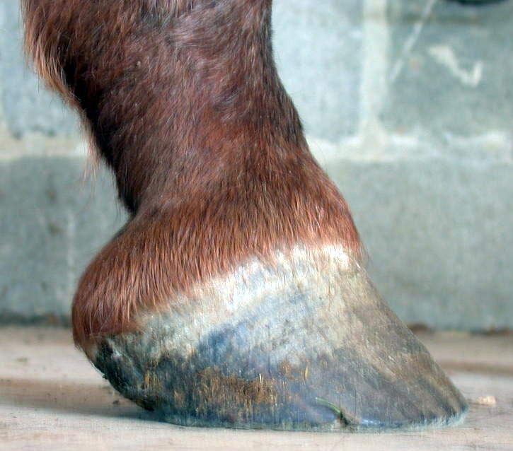
This 20 yo OTTB, who went barefoot at the age of 15 after a fatal navicular diagnosis, completed the challenging hunter pace completely bare (no hoof boots). The terrain included dirt roads, hard-packed grass fields after weeks of no rain, softer grassy fields, a ditch and muddy bank on the edge of a stream, a good bit of rocky going, asphalt road crossings, and deep plowed up cornfields.

Besides the beautiful red ribbon (visible on the bridle above), the prize was one free hunt capping fee for the ’06-’07 Season.  Barefoot fox-hunting next?

The worst thing that happened was that the rider lost her favorite crop which had been in her possession for many, many years and miles, was just the right length and balance, and probably cannot be replaced.

One month later horse and rider completed another hunter pace (Spring Valley Hounds, New Vernon, NJ), this time in the Open Division.  Again they rode shoe and boot-less and although the footing was more forgiving, there were 2’6 high jumps, a much faster pace (thanks to improved fitness) and stiffer competition.

Here is a picture of the pair jumping a stone wall at Spring Valley Hounds.

His complete case will be published in future posts, so check back!

Splints

The horse, a 5 yo Clydesdale/TB cross, was shod for a period of about six months (or less) while four years old, during which time he developed splints.

After shoes were removed, he was trimmed for minor lateral imbalances, pigeon toes, slightly high heels, and too long quarter walls resulting in wavy hoofwall rings. The splints were reduced in size as a side effect of that trimming, leading one to conclude there is a possibility that pigeon toed “conformation”, combined with concussion, can cause splints.

splintpu.jpg

Approx. time of this trim July ’06.

Continue reading

Quarter Crack

 The horse, about 7 or 8 years old had been barefoot for most of his life, with apparently appropriately short heels and toes, but still had persistent quarter cracks on both feet. His feet were badly unbalanced and had underrun heels. 

 qcrack.jpg

Right Front Before

during-crack.jpg                                          sole-heels.jpg

During                                                              Before Sole View RF foot

Heel position improved, removing lever forces on hoofwall. Hoof/Pastern Axis improved. The crack is still visible but is no longer splitting.

The sole view shows uneven underrun heels – the lateral heel of the RF is more underrun than the medial one and is further forward underneath the foot (refer to yellow line and arrows) , which is contributing to the quarter crack on that side of the foot. During the following months the heels were brought back to the widest point of the frog where they lined up with each other.

Continue reading

The Heel Pain Stance and Proper Body Posture

 An example of incorrect stance due to heel pain or imbalance:

beforelillybody1.jpg

This horse exhibits signs of heel pain by placing its left leg behind its right one, behind the vertical.  The neck is held stiffly in an upright position with the bottom of the neck appearing “longer” than the top and rigidity in the lower muscles.  The triangle at the withers and scapula show depressions in the muscles. When holding its body this way for extended durations, the underneath neck muscles become overdeveloped and the topline underdeveloped. The ears are grumpily flattened.

A horse is standing correctly when its cannon bones are vertical (or perpendicular to the ground). If a horse continually stands with one or both legs behind the vertical, he likely is experiencing heel pain. rfbef.JPG It can also be a matter of unbalanced feet, wtih the heels on one foot being higher than the other. The higher-heeled foot tends to be the one held back as the horse seeks to balance himself. Sometimes it can be difficult to distinguish between heel pain, and a horse not weighting its heel from long term joint adaptation.


The pony stands up correctly after a balanced trim  rfaftsm.jpg

Continue reading

High Heels and Other Issues

This is a case of a 14 yo QH with an improperly healed fracture of a large left hind leg bone which prevented him from using the leg properly and moving or standing comfortably. At the time he was reclaimed by his owner, he walked with great difficulty.

stifle2.JPG

The injury to the LH is apparent in his odd stance. It prevented him from holding his foot up for trimming and thus all his feet had become overgrown.  He was unable to hold up his feet or bend his legs in any way that allowed the toes to be trimmed, and no sling was available. Consequently, his toes were not shortened enough during his initial trims seen here.

Hind Feet

Typically, high heels are more common in the front feet than the hinds.  Here the hind heels became high as a result of the injury.

lh1.JPG  lh2.jpg  lh3.jpg

The Left Hind foot prior to trimming, and during the course of the next six months.

Continue reading

High Heels I (High – Low Syndrome)

High/Low Syndrome

highlow.jpg

This horse exhibits what is sometimes called “high/low syndrome” with one foot having high heels usually on a contracted foot (which may or may not be a clubfoot),  and the other with low and sometimes underrun heels,  flat and  lacking concavity.  The horse usually stands in a scissor stance, with the high-heeled foot back behind him and the flat foot in front of him, due to the discomfort of weighting the back of the high-heeled foot, which creates a vicious cycle of exacerbating the high heeled condition.

lfaug04bef.jpg               redaug04-2.JPG     9904side-2.JPG   112404lfside2.JPG

Before                                      August                          September                         November

Continue reading

Homebred TB Yearling. Over at the Knee

This TB colt had been described as being over at the knee, or tied-in, and that this was genetic and conformational and therefore nothing could be done about it. After his first trim  (at about 1 yr old) he stood with both front legs straight.  In the photo below at 1.5 yrs, (one month into the trim, with long bars) he is standing with cannon bones slightly behind the vertical.  Right after being trimmed, he stands more squarely.  His conformation critique also stated his pasterns were a little long.  The angle and therefore the apparent length, has been improved by trimming  (photo 8).

   At 6 mos.                                                                    At 1.5 yrs.

 stupx0.JPG                                 willy-upx6.JPG                b2oct2006.JPG

  Before barefoot trim                          One month after trim            Right after a trim

 The TB gelding, bred for the track, was born in Mar ’05. He was receiving a farrier pasture trim during his first year which rasped the toes handsomely but removed no sole, or bar. His heels were reasonably low. The young gelding lives in an ideal environment: 24/7 turnout in a rural area 100 miles north of New York City, in a herd in large pasture with a natural water source and on a variety of terrains, mostly hard and rocky.

At about one year old, he was becoming very inactive for a TB colt, standing around a lot and not getting much movement. The owner was concerned he might have contracted Lyme’s disease and had the vet run tests which were negative, thus, the vet conlcuded it was “just the way he was”.  Soon after he received his first correct barefoot trim which included removing very large amounts of severely overgrown bar (about twice what is shown in photo 4), lowering the heels, and shaping the toe. Immediately after the trim he ran off bucking, at a gallop and to the great relief of the owner.

                 May ’06                                      Oct ’06

LF     1.  lfsafx1.JPG              2. lfsafx7.JPG                             

RF     3.  rfsafx3.JPG            4.  rfsbf-x8.JPG    5.   rfsafx9.JPG          

                                                                         before trim               after trim

 LH      6. lhsafx4.JPG          7 .   lhsafx10.JPG


LF        8.  lfsideafx11.JPG

 Six weeks after the first trim the foot appeared to double in width, more than what could be attributed to normal growth. The long and deep bar seemed to be impinging the growth of the foot. Now, the coffin bone will be able to develop to its full genetic potential. There is also a significant increase in width visible in the six months from May ’06 to Oct ’06 (compare 1. to 2. and 3. to 5.)

The hind foot also shows correct development, in that it is not becoming as wide, but the spade shape of a hind foot is becoming more apparent as the horse develops (6. to 7.)

Clubfoot

rfsidebef.JPG

side2.JPG               sole-2.JPG

This is a 5 yo Morgan mare. The trim was started at age 3, and the second row of pictures shows the progress after about one year.

With frequent trimming, the heel has been kept low and the coffin bone placed in a near-ground parallel orientation.  This has enabled the dish in the toe wall to grow out, and the hoof rings to become somewhat more concentric (more equal distance between them at the heel and at the toe.  When the heel grows faster than the toe, the rings are wider at the heel.   A healthy foot’s growth should be even all around with equidistant rings).  However by the age of three there was already considerable joint adaptation in the coffin joint, creating the broken forward hoof-pastern axis. 

dressage-test-corner.jpg © James Matteson

The mare recently competed in her debut dressage show, winning her Intro test amongst a class of six, as well as getting an award for second highest score of the day.  She has a bright future ahead of her as an all-around fun horse.

No More Ringbone

A Horse Who No Longer Has Ringbone

051005before.JPG

Before

She is a 20+ yo Morgan mare who was grade 5 lame and retired.  The original diagnosis for the lameness was navicular. With the prescribed bar shoes, she pulled a sesamoidean ligament within 24 hours. The shoes were removed, and a second opinion diagnosed the ringbone. The treatment prescribed was retirement, with the advice to give bute the day before if the owner wished to ride her. 

Within seven months of correct trimming she has regained complete soundness and gets regularly ridden.

rfside.JPG

After

The long toe was  later shortened.

Improvement in the quality and appearance of the coat is visible (not so coarse, has more shine), as a result of the increased circulation to the hooves and legs.

The lateral cartilage is no longer so prominent and pushed up out of the hoof capsule, now that the heels have been brought back even with the widest part of the frog, and the walls in the quarters have been shortened. Such a displacement of the lateral cartilages can lead to ossifications such as sidebone, or contribute to ringbone. Most importantly the steep pastern has taken on a better angle by virtue of the heels being lowered and brought back even with the widest part of the frog. This steep alignment is the factor that most contributes to and predisposes a horse to developing ringbone, as it puts excess and unnatural strain on the extensor tendon; its attachment  point becomes inflamed and eventually stabilizes by calcifying.